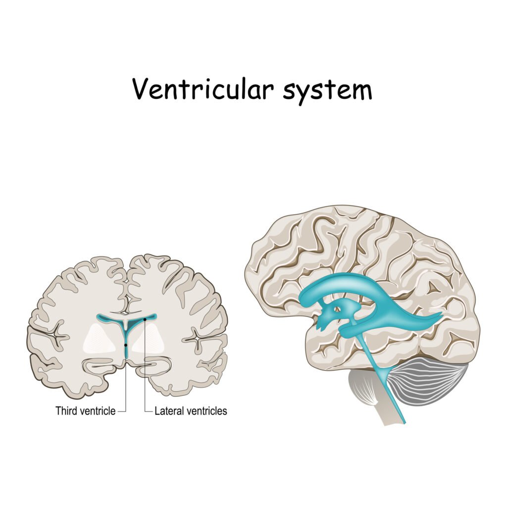
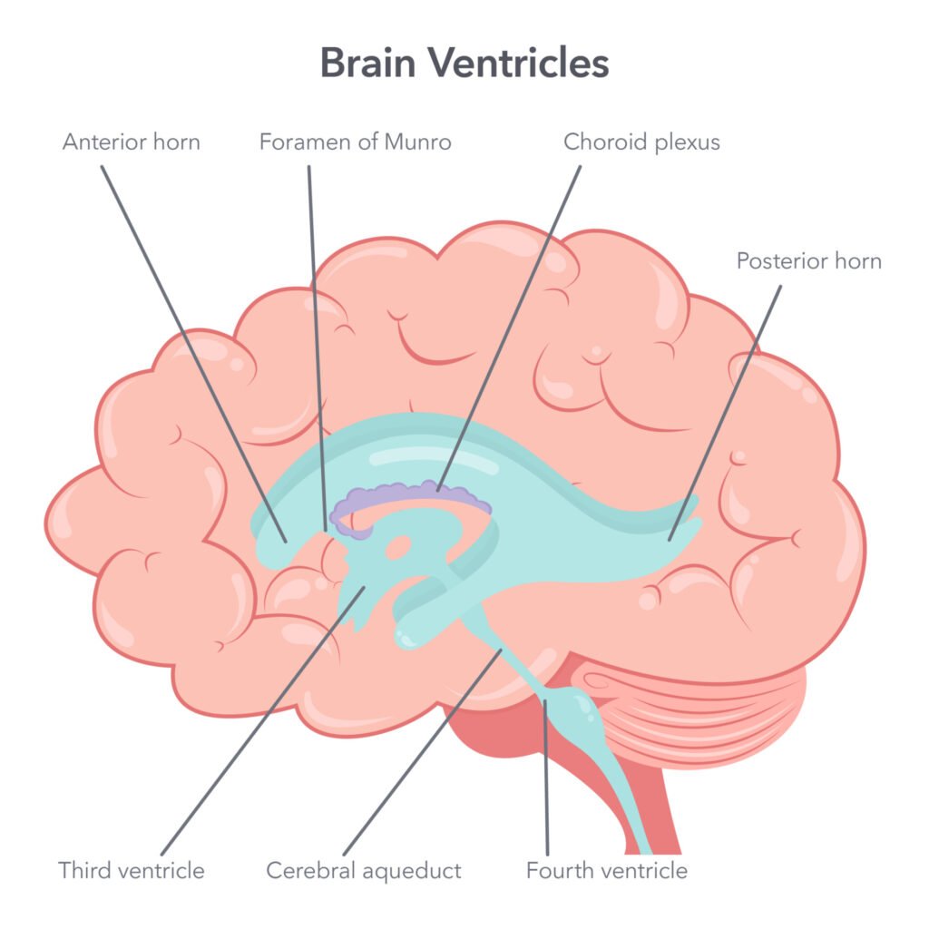
The ventricular system is a network of fluid-filled cavities within the brain, including the lateral, third, and fourth ventricles, which produce and circulate cerebrospinal fluid (CSF). CSF provides cushioning, nutrients, and waste removal for the brain, helping maintain a stable environment for optimal neural function.
Disruptions in the ventricular system can lead to neurological disorders and conditions, emphasizing its crucial role in brain health.
The types of ventricles
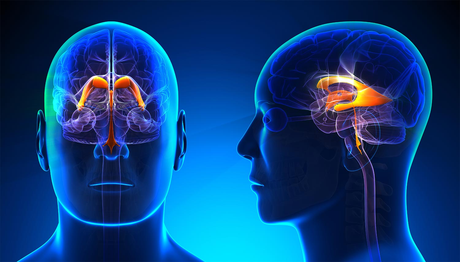
The ventricles of the brain are cavities which produce and store a substance known as cerebrospinal fluid (CSF). The brain’s ventricular system is comprised of four ventricles as well as small structures which connect each ventricle called foramina.
In this way, the ventricles are linked to each other to function as they should. The ventricles are essential for maintaining the central nervous system (CNS).
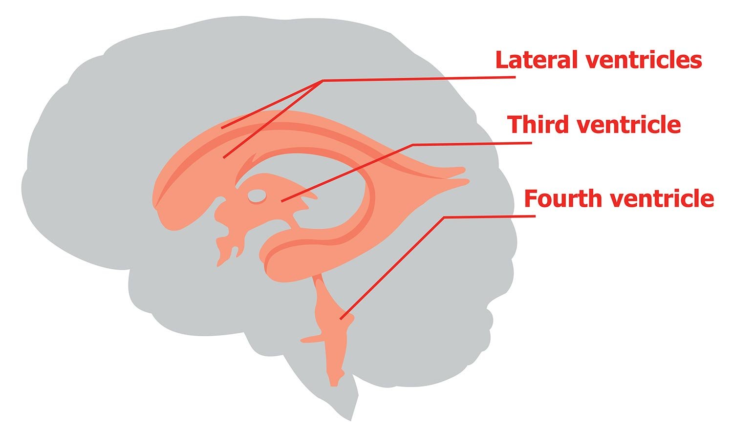
There are four ventricles of the brain: the 2 lateral ventricles, third ventricle, and fourth ventricle. The ventricles are lined with a specialized membrane called the choroid plexus, which is made up of ependymal cells.
Ependymal cells are glial cells, types of cells which provide physical and metabolic support to neurons.
These specific cells are tailored to produce cerebrospinal fluid (CSF) and they secrete the fluid into the ventricles at a relatively constant rate. Each brain ventricle’s main function is to produce, secrete, and convey CSF.
Lateral ventricles
The first and the second ventricles are known as the lateral ventricles. Each of the lateral ventricles are located in each hemisphere of the cerebral cortex, one in the left hemisphere, one in the right.
The lateral ventricles are C-shaped structures and have three horns which project into three lobes of the brain: the frontal, occipital, and temporal lobes.
The lateral ventricles are connected to the following ventricles, the third ventricle, by an opening called the interventricular foramen (or foramen of Monro). The volume of the lateral ventricles is also seen to increase with age.
Third ventricle
The third ventricle is a very narrow, funnel-shaped structure situated between the right and left thalamus, in the midline between the right and left lateral ventricles, just above the brain stem.
As well as producing CSF, the third ventricle has direct communication with each lateral ventricle through the Monro foramen.
This ventricle also interacts with the fourth ventricle through what is known as the cerebral (or Sylvius) aqueduct, a channel that allows CSF to pass between the third and fourth ventricles.
Fourth ventricle
The fourth ventricle is the last ventricle of the system which receives CSF from the third ventricle via the cerebral aqueduct.
The fourth ventricle is a diamond-shaped structure which lies within the brain stem, at the junction between the pons and the medulla oblongata. It has four openings through which CSF drains into two places of the brain.
CSF drains into the central spinal canal, bathing the spinal cord. It also drains into the subarachnoid cisterns, bathing the brain, between the arachnoid mater and pia mater. Here the CSF is reabsorbed back into the circulation.
Cerebrospinal fluid (CSF)
The human brain is fully encased in a skull to protect it from damage. For further protection, the brain is covered in three meningeal layers: pia mater, arachnoid mater, and dura mater.
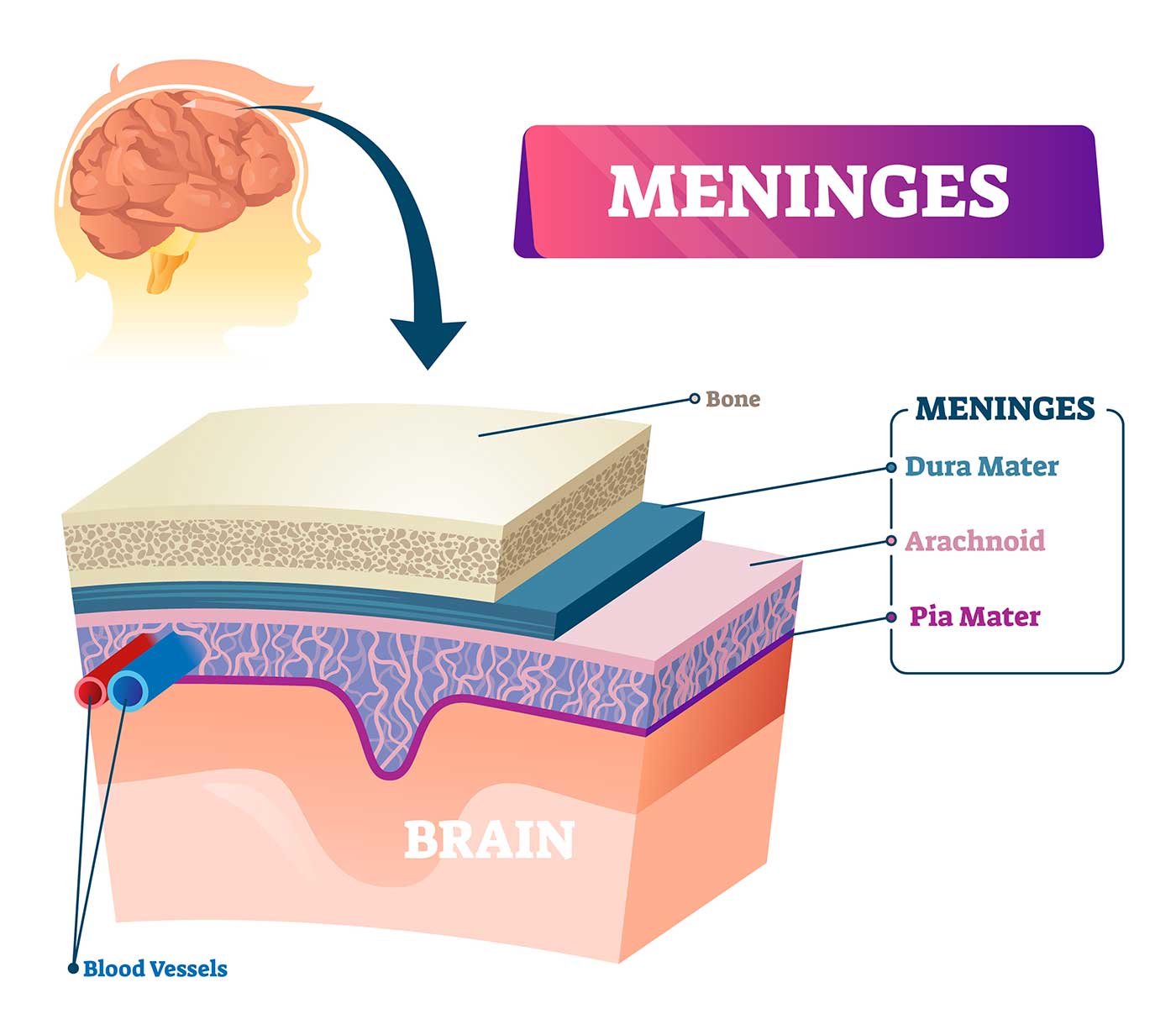
Pia mater is a delicate inner layer, the arachnoid mater is the middle layer consisting of a web-like structure, and the dura mater is the tough outer layer.
Despite all this protection, there is still space surrounding the brain which can make it vulnerable to injury. This space is therefore occupied by CSF, which is produced by the ventricular system.
It wasn’t until 1794 that it was discovered that the ventricles were filled with CSF.
The main purpose of the ventricular system is to produce CSF which serves many functions, but mostly to protect the CNS. Aside from the CSF, the ventricles are hollow.
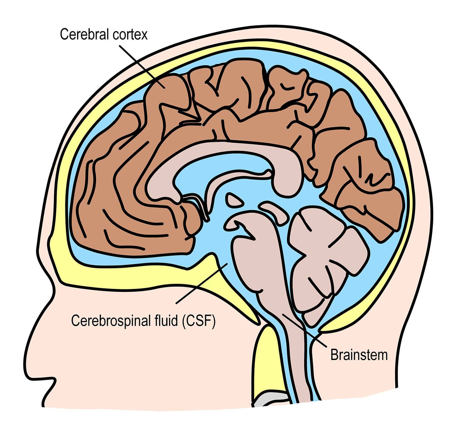
Cerebrospinal fluid (CSF) is constantly bathing the brain and spinal column. It is produced by the choroid plexus, located in the living of the ventricles.
Plasma is filtered from the blood to produce CSF. In this way, the exact chemical composition of the fluid can be controlled.
About half a litre of CSF is produced every day by the ventricles and flows through these ventricles in a constant stream.
Cerebrospinal fluid functions
-
Shock absorption – the CSF encasing the brain absorbs the shock of trauma to the head and spine so that the brain does not collide with the skull.
Through cushioning the brain, it helps to protect against potential injuries that may be caused by pressure or force.
-
Making the brain buoyant – CSF makes the brain buoyant, reducing the physical stress it would otherwise experience from the forces of gravity and movement.
In fact, without being suspended in the fluid of some kind, the brain can become distorted under its own weight and the delicate tissue can tear.
-
Nutrition – the CSF provides the central nervous system with essential nutrients such as glucose, proteins, lipids, and electrolytes, all to keep it healthy and functioning properly.
-
Waste removal – the CSF is responsible for cleaning through the subarachnoid space of the spine, cleaning up toxins and waste products that are released by nerve cells .
This waste is then carried to the lymphatic ducts for filtration. The toxins are emptied into the bloodstream where they are eventually removed by mechanisms such as kidney filtration.
-
Intracranial pressure – a steady flow of CSF circulating around the brain keeps it stable. If there were too much CSF, as a result of a traumatic brain injury or brain tumor for instance, this can raise the intracranial pressure.
-
Temperature – because of the circulation of CSF, the temperature of the brain and spine remains stable.
-
Immune function – the CSF contains numerous immune cells which work to monitor the central nervous system for foreign agents that could potentially damage the vital organs.
The flow of cerebrospinal fluid
Once produced in the lateral ventricles, CSF fills the cavities then proceeds to leave via the interventricular foramen to enter the third ventricle.
In addition to the CSF being produced in the lateral ventricles, the CSF is also produced in the third ventricle and then exits the space through the cerebral aqueducts to enter the fourth ventricle.
Very little CSF is produced in the fourth ventricle. However, it, along with that coming from the previous ventricles, exits the fourth ventricle to enter either the central canal of the spinal cord or into the subarachnoid cisterns, spaces formed by openings in the subarachnoid space in the meninges of the brain.
Associated conditions
Conditions associated with ventricular system dysfunction can be commonly attributed to infections, head trauma, and bleeding in the brain.
The different causes can lead to inflammation in the ventricles. Inflammation can block the flow of CSF, causing the ventricles to swell in size and placing pressure on the brain.
The following ventricle-related conditions can be life-threatening:
Hydrocephalus
The rate of CSF production in the ventricles is constant regardless of changes in pressure. This can become a problem if the passage of CSF is blocked somewhere within the system.
CSF will continue to be produced, but it will have no means of exiting the system. This can cause pressure within the ventricles to increase, and the rising pressure may eventually force the ventricles to expand.
The expanding ventricles can then impose on other brain structures causing a variety of complications. When this occurs in children whose skull has not completely hardened, it can cause the head itself to enlarge. This condition is known as hydrocephalus, also known as ‘water on the brain’.
This condition can be present at birth due to a genetic or developmental abnormality, but it can also develop due to a brain or spinal cord tumor, stroke, head trauma that causes bleeding in the brain, or an infection.
There are two primary types of hydrocephalus: communicating and non-communicating hydrocephalus. Communicating hydrocephalus is when the CSF becomes blocked in the subarachnoid space after it exits the ventricles.
The non-communicating hydrocephalus however, is when the CSF becomes blocked in one or more structures that connect the ventricles.
Whilst this condition is more common in infants, it is also common in adults over the age of 60 years old.
The symptoms of hydrocephalus in infants include:
-
The head growing in size at a rapid pace
-
Trouble feeding
-
The soft spot on their head bulging
-
Sleepiness
-
Irritability
-
Seizures
The symptoms of hydrocephalus in older adults include:
-
Difficulty walking, balancing, or lifting their feet
-
Rapid onset of dementia or other cognitive impairments
-
The inability to hold their bladder
Hydrocephalus can also occur in all other age groups, although not as common. Some of the symptoms include:
-
Headache
-
Changes in vision
-
Difficulty walking or talking
-
Personality changes
-
Memory loss
Meningitis
The meninges are membranes lined in the subarachnoid space. Meningitis is a condition which can develop when this lining, along with the CSF becomes infected and inflamed.
Some possible causes of this are bacterial, viral, parasitic, or fungal infections. The most serious type of infection being bacterial meningitis.
The bacterial type of meningitis can block the flow of CSF in the subarachnoid space and in the ventricles. This then will cause hydrocephalus.
The symptoms of meningitis tend to come on rapidly and can include:
-
Headaches
-
Fever and chills
-
Stiff neck
-
Sensitivity to light
-
Nausea or vomiting
-
Confusion
-
Seizures
Ventriculitis
The choroid plexus in the ventricles contains a layer of ependymal cells, which forms the ependymal lining.
Ventriculitis occurs when this lining becomes inflamed due to meningitis, head trauma, or as a complication of brain surgery.
The symptoms of ventriculitis can mimic those of meningitis and can include:
-
Headache
-
Fever and chills
-
Stiff neck
-
Confusion
-
Seizures
Brain hemorrhage
Injuries that cause bleeding in the brain are known as hemorrhages. Injuries such as a stroke, ruptured aneurysm, or traumatic brain injury can cause bleeding, specifically in the subarachnoid space or the ventricles.
These are known as subarachnoid or intraventricular hemorrhages. Both types can result in hydrocephalus as the blood clots form and cause a blockage in the flow of CSF in and around the brain ventricles.
Symptoms of brain hemorrhages can come on suddenly and can include the following symptoms:
-
Severe headaches
-
Blurred or double vision
-
Stiff neck
-
Slurred speech
-
Sensitivity to light
-
Weakness on one side of the body
-
Nausea or vomiting
-
Loss of consciousness
References
Neuroscientifically Challenged. (2015, February 1.). Know Your Brain: Ventricles. https://neuroscientificallychallenged.com/posts/know-your-brain-ventricles
Crumble, L. (2021, October 28). Ventricles of the brain. Kenhub. https://www.kenhub.com/en/library/anatomy/ventricular-system-of-the-brain
Vega, J. (2021, October 21). The Anatomy of the Brain Ventricles. Very Well Health. https://www.verywellhealth.com/brain-ventricles-3146168
Jones, O. (2020, November 13). The Ventricles of the Brain. Teach Me Anatomy. https://teachmeanatomy.info/neuroanatomy/vessels/ventricles/
