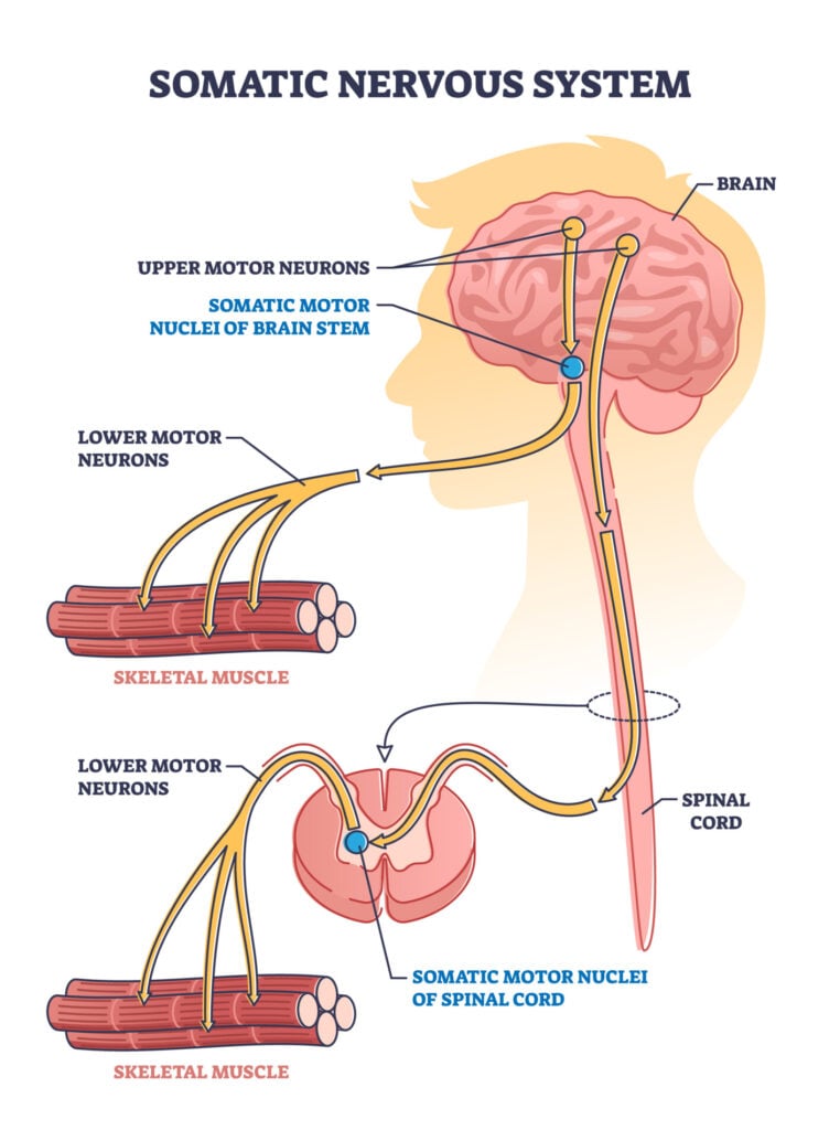
The somatic nervous system (SNS) is a component of the peripheral nervous system responsible for voluntary movement and the reception of external stimuli. It regulates activities under conscious control, such as skeletal muscle contraction. The SNS comprises motor neurons that activate muscles and sensory neurons that relay information from sensory receptors to the central nervous system.
Take-home Messages
- The somatic nervous system (SNS) is part of the peripheral nervous system (PNS) and is associated with activities traditionally thought of as conscious or voluntary, such as walking.
- The somatic nervous system consists of motor neurons and sensory neurons, which respectively transmit motor and sensory signals to and from the central nervous system (CNS).
- The somatic nervous system controls voluntary movements, transmits and receives sensory information (e.g., sight, taste, touch), and is involved in reflex actions without the involvement of the CNS so that the reflex can occur very quickly.
The somatic nervous system (SNS) plays an important role in initiating and controlling nearly all voluntary movements of the body. The SNS is a branch of the peripheral nervous system, along with the autonomic system (ANS), although they function in different ways.
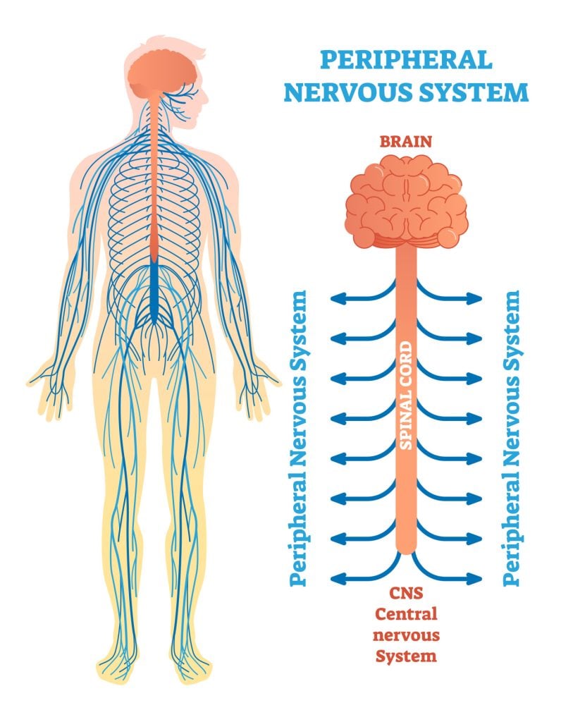
Whilst the autonomic nervous system (ANS) regulates automatic behaviors, such as breathing and heart rate, which do not require conscious thought, the somatic nervous system (SNS) mostly regulates conscious body movements.
The main function of the SNS is to transmit signals between the body’s muscles and the brain and the central nervous system (brain and spinal cord) to control voluntary movement and reflexes.
It can do this by processing sensory information, which arrives through external stimuli via the senses (sight, taste, touch, smell, and hearing).
Somatic nerves also react to the environment through somatosensory and motor functions. For instance, when humans or animals are experiencing coldness (somatosensory function), they can move (motor function) to a warmer place for survival.
Being able to control the internal environment is called homeostasis (“homo-” meaning same and “-stasis” meaning state of equilibrium), which needs to be maintained for survival. The SNS and the ANS work together to regulate bodily function and provide reactions to external stimuli (Fukudo, 2012).
The SNS consists of neurons (nerve cells) located in the brain stem, spinal cord, and dorsal root ganglia.
They are extremely long as they do not synapse until they reach their termination point at the skeletal muscle (Rea, 2014).
Somatic Nervous System Function
What does the somatic nervous system do?
The main function of the SNS is to control all voluntary movements. There are receptors in the skin and sense organs (e.g., eyes, mouth, nose, and ears) which are able to detect changes in the environment, such as temperature, light, pressure, or texture.
Once environmental changes have been detected, impulses are created within the sensory neurons, which then carry signals through the spinal nerves to the spinal cord. These signals will then travel up the spinal cord to the brain.
The brain will then integrate this sensory information and determine an appropriate response. This response is then transmitted to the spinal cord, reaching motor neurons.
The impulses will then be carried through the motor neurons out of the spinal cord and continue to the skeletal muscles, causing them to contract if needed.
Essentially, there are two pathways involved in the SNS. The afferent pathway will carry sensory information from sensory organs to the CNS. In contrast, the efferent pathway will carry motor information from the CNS to skeletal muscles to regulate motor functions.
Reflex Arcs
As well as controlling all voluntary muscular systems of the body, the SNS also processes muscle reflex arcs. Reflex arcs are neural pathways that produce involuntary movements, typically in response to stimuli perceived as imminent danger.
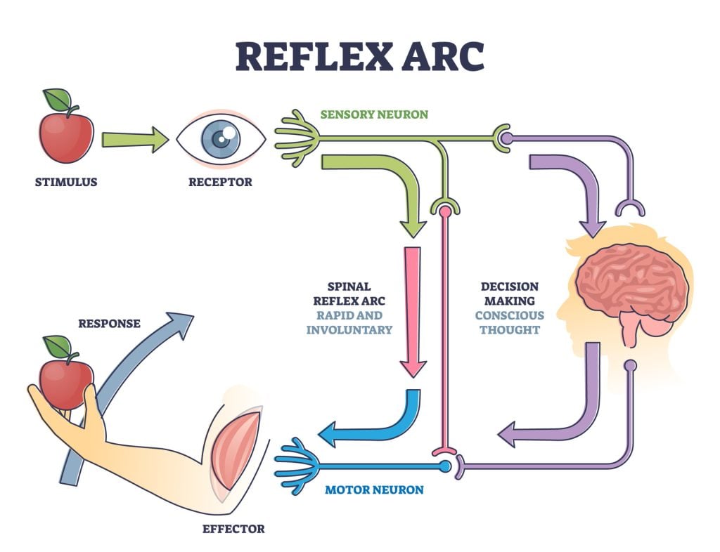
This occurs when sensory neurons sense something within the environment and carry this signal directly to the spinal cord, but this is not transmitted to the brain.
The spinal cord will instead transmit signals through the motor neurons to the muscles in order to trigger a reflex movement. This way, the muscles move without any input from the brain to generate a response that is so fast that it is completed almost automatically.
An example of a reflex arc being used would be when moving a hand away after touching a hot surface. Another instance is the ‘knee jerk’ reaction.
Parts of the SNS
The SNS consists of two major types of neurons; sensory and motor neurons. These neurons function to transmit signals throughout the body.
Neurons contain an axon, which is the longest part of the cell, enabling signals to be transmitted through them.
There are also dendrites branching off the neuron soma (cell body) or at the neuron endings, which can make connections with, or synapse, other cells’ axons to receive input.
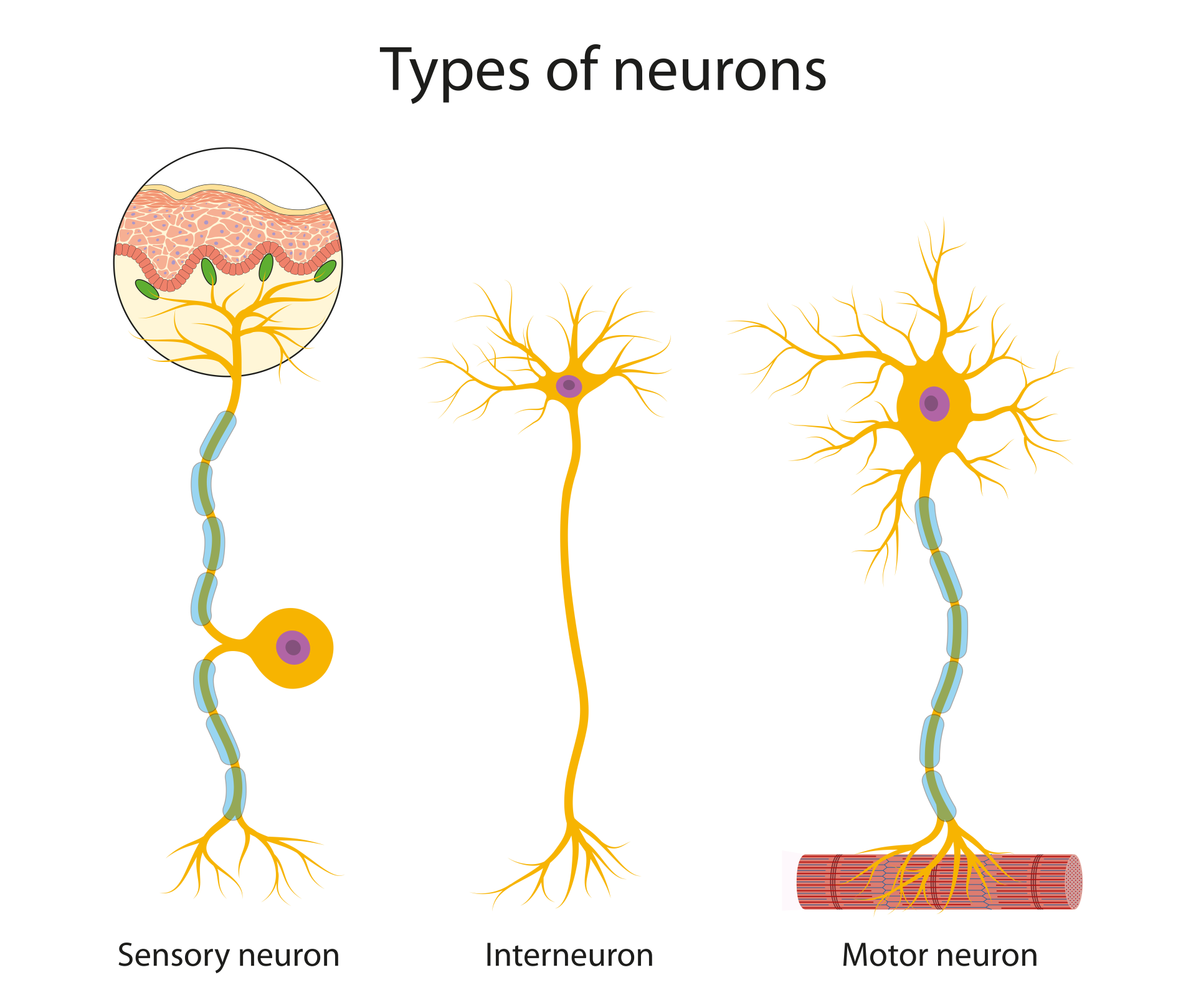
Sensory neurons within the SNS, also known as afferent neurons, carry sensory information from the body to the central nervous system (CNS).
Sensory neurons of the SNS send information to the central nervous system from external stimuli from the senses, such as information about the texture of an object.
Motor neurons within the SNS, also known as efferent neurons, carry motor information from the CNS to muscle fibers throughout the body.
These types of neurons transmit signals from the spinal cord and brainstem to skeletal muscle to directly control muscle movements.
The contraction of a muscle is caused by the activation of motor neurons that supply the skeletal muscle. The connection between motor neuron and skeletal muscle is known as the neuromuscular junction (NMJ). Acetylcholine is the primary neurotransmitter (chemical messenger) transmitted through the NMJ.
Whilst acetylcholine has an inhibitory effect in the ANS, within the SNS, it is excitatory. The electrical signals from the motor neurons are converted to chemical messages at the NMJ, which is where acetylcholine is released by the motor neuron (Cuevas, 2015).
Cranial nerves, which originate primarily in the brainstem, play a vital role in carrying information to and from the brain. These nerves allow sensory information to transmit from the organs of the brain (ears, eyes, nose, and mouth), as well as transmitting motor information from the brain to these organs.
For instance, when eating food, the brain will convey motor messages through the nerves to move the mouth in order to chew and swallow.
Of the twelve cranial nerves, ten originate from the brain stem and primarily control the voluntary movement and structures of the head.
While cranial nerves are responsible for head and neck sensation and movement, spinal nerves are responsible for bodily sensation and movement. Spinal nerves send somatosensory information into the spinal cord. Then, they send motor information back out of the spinal cord.
These signals are then transmitted to the brain for processing. Spinal nerves control the function and movement of the rest of the body below the head and neck (Akinrodoya & Lui, 2020).
Damage
As the SNS is responsible for receiving sensory information and motor movements, symptoms associated with damage to this system are experiencing numbness, muscle weakness, and pain.
Diabetes is the most common cause of neuropathy in the PNS, but damage can also be the result of autoimmune diseases, infections, and trauma. Damage to the nerves through injury can affect the functions of the afferent and efferent pathways of the SNS.
A number of diseases involved in sensory and motor control arise from either an issue with the CNS, PNS, or the muscle itself. Due to the range of functions covered by the SNS, these diseases can either be localized to a specific region of the body or it can be widespread and generalized.
Some conditions could be the result of issues with the axons of the neurons (axonal neuropathy) or could be the result of issues with the myelin sheath (demyelinating neuropathy), which is the protective layer of the neurons (Akinrodoya & Lui, 2020).
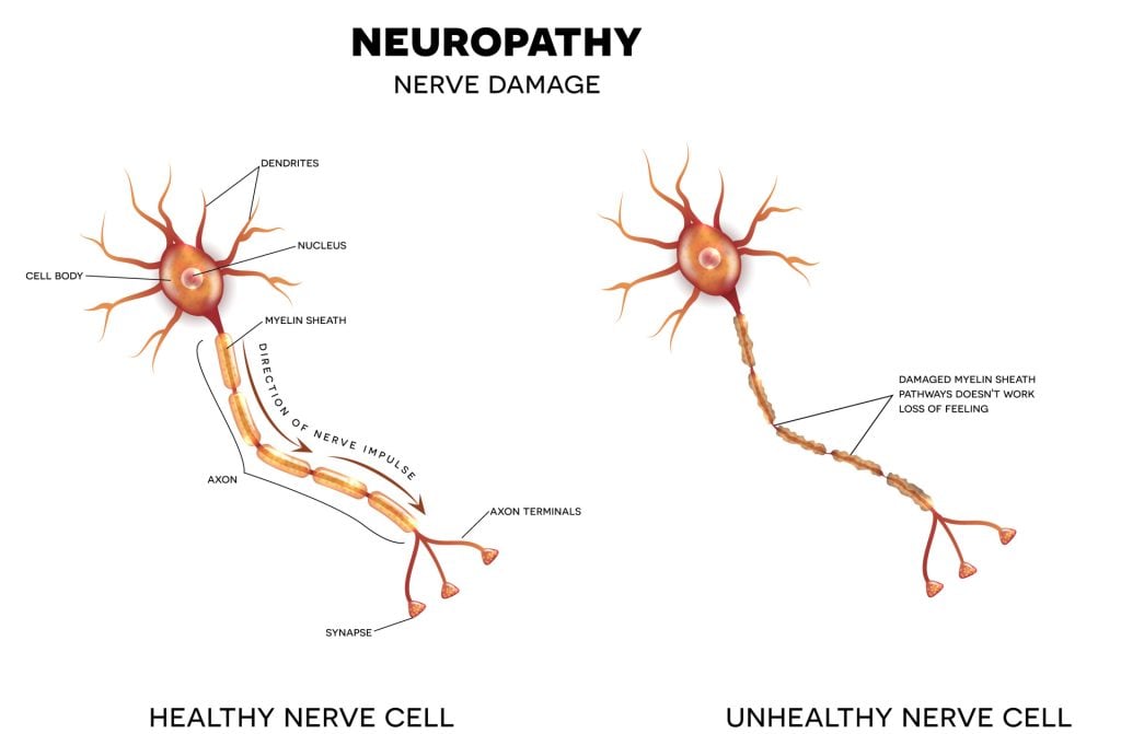
Motor neuron disease is the result of the death of neurons, making it a neurodegenerative disease. Eventually, the muscles of people with this disease will deteriorate, resulting in a loss of function.
Multiple sclerosis is an autoimmune disease that causes the destruction of peripheral nerves, resulting in a variety of sensory and motor problems.
Whilst these diseases are not always preventable, there are some lifestyle changes that can aid in stopping the preventable symptoms of SNS damage/weakening.
This can include sticking to a healthy diet in order to maintain a healthy weight, not smoking, avoiding alcohol, regular exercise, and correcting vitamin deficiencies to limit the chance of developing issues.
References
Akinrodoye MA, Lui F. Neuroanatomy, somatic nervous system. StatPearls [Internet]. Treasure Island (FL): StatPearls Publishing. Updated April 2, 2020.
Cuevas, J. (2015). The Somatic Nervous System . Reference Module in Biomedical Sciences. Elsevier.
Dorland, W. A. N. (2011). Dorland’s Illustrated Medical Dictionary E-Book. Elsevier Health Sciences.
Fukudo, S. (2012). Hypothalamic-pituitary-adrenal axis in gastrointestinal physiology. In Physiology of the gastrointestinal tract (pp. 791-816). Elsevier Inc.
Rea, P. (2014). Introduction to the nervous system. Clinical Anatomy of the Cranial Nerves; Rea, P., Ed.; Academic Press: Cambridge, MA, USA.
